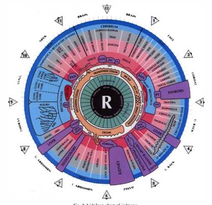
Live Blood Cell Analysis

The test is carried out by obtaining a drop of blood from the finger tip using a sterile lancet.

The blood is placed on a microscope slide, covered with a coverslip to prevent drying, and immediately observed under the microscope. Darkfield microscopy allows an almost three dimensional look at the blood. The blood is magnified up to 1000 times. A video camera is hooked up to the microscope so that you may watch the entire procedure on a monitor.

You will be able to see the activity of your blood on the screen, the red blood cells, white blood cells, plasma and what is floating in the plasma, like bacteria, yeast, crystals, plaque, and more. The blood is observed for 30 minutes. Once the blood leaves your body it begins to die. How quickly the morphological changes happen can indicate your present health condition.

This method of analysis is different from the usual laboratory tests that quantify the levels of certain components in a sample of blood. Live blood screening gives an indication as to the QUALITY of an individual’s blood – an important foundation of preventative health care showing the effects that diet, lifestyle and stress can have on our well-being

Normal Blood
Abnormal Blood

Depending on the irregularities found in the blood, the Live Blood Cell Analysis can help to reveal the following:

  • Immune System Function
  • Free Radical Cellular Damage
  • Cell Size & Shape Abnormalities
  • Early Recognition of Cardiovascular Risk Factors
  • Essential Fatty Acid Deficiencies
  • Vitamin & Mineral Deficiencies
  • Digestive Disorders
  • Low Iron/Anaemia
  • Atherosclerotic Plaque
  • Allergies Tendencies
  • Hormonal Imbalances
  • Uric Acid Crystal & Risk for Gout
  • Kidney/Liver/Spleen Stress
  • Chemical/Heavy Metal Toxicity
  • Degenerative Disorders
  • Parasites
  • Dehydration
  • Poor Blood Circulation
  • Clotting Abnormalities
  • Bacterial Imbalances
  • Yeast/Fungal Tendencies


Dry Blood Cell Analysis

The Dry Blood Analysis or procedure involves puncturing a client’s fingertip and allowing a bead of blood to form. This is left on the finger for 30 seconds to allow the clotting cascade to develop. A microscope slide is then pressed onto the drop of blood eight times. The slide is left to dry, which takes about 15 minutes, and in that time the eight blood layers will go through an extraordinary transformation.

Dry Blood Analysis can help :

  • Detect the presence of inflammation
  • Assess the progression of inflammatory processes – chronic or acute
  • Determine the location of the inflammatory process – organ, tissue or system specific
  • Monitor the progression or reduction of the inflammatory process
  • Detect heavy metal toxicity
  • Reveal the presence of parasites
  • Show mineral and vitamin imbalances
  • Show antibiotic damage
  • Detect bowel inflammation
  • Assess the lymphatic burden
  • Assess protein metabolism
  • Reveal Thyroid/Endocrine imbalances